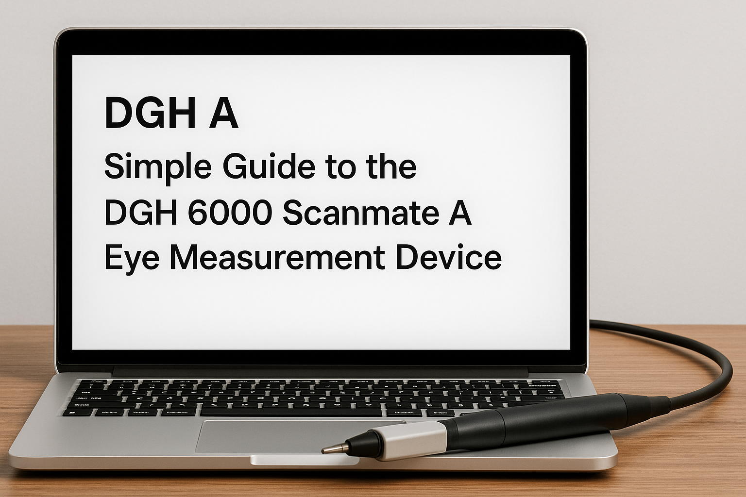
DGH A: A Simple Guide to the DGH 6000 Scanmate A Eye Measurement Device
The human eye is small but very complex. Every tiny part of it helps us see the world clearly. When doctors perform eye surgery, such as cataract or lens replacement surgery, they need to know the exact size and shape of the eye Insetprag
A few millimeters can make a big difference in choosing the right artificial lens for the patient. To make these measurements accurate, eye doctors use a special ultrasound machine called DGH A, or the DGH 6000 Scanmate A.
This device measures how long the eye is and how deep the front parts are. The information helps doctors plan safe and successful eye surgeries. This article explains everything about DGH A — what it is, how it works, its parts, how to use it, and why it is helpful in eye care.
What Is DGH A
DGH A (also known as DGH 6000 Scanmate A) is a biometric ultrasound device used to measure the internal structure of the eye. It uses sound waves to create a simple line graph, called an A-scan, showing where the main parts of the eye are located.
Doctors use this information to:
-
Measure axial length — the distance from the front of the cornea to the retina.
-
Measure anterior chamber depth — the space between the cornea and the lens.
-
Measure lens thickness — how thick the eye’s natural lens is.
These numbers help the doctor calculate the intraocular lens (IOL) power needed for cataract surgery or other eye procedures.
DGH A is small, portable, and connects easily to a computer. It is made by DGH Technology Inc., a company that makes professional eye ultrasound equipment used around the world.
How DGH A Works
The DGH A works using ultrasound — high-frequency sound waves that can travel through soft tissue.
Here’s what happens step by step:
-
The probe sends a short pulse of sound into the eye.
-
The sound waves bounce back (echo) from different parts of the eye.
-
The machine measures how long the sound took to come back.
-
The software turns that time into distance, showing how far each layer is.
The result appears as a waveform on the screen — a line with small peaks that show the cornea, lens, and retina.
The DGH A can be used in two ways:
-
Contact mode: The probe gently touches the cornea.
-
Immersion mode: The probe stays just above the cornea with a thin layer of fluid in between, avoiding pressure on the eye.
Main Features of DGH A
The DGH 6000 Scanmate A includes several helpful features that make it easy for clinics to use.
Features:
-
Real-time guidance: The software gives a sound and visual signal to help the user keep the probe aligned correctly.
-
Compression lockout: Prevents readings if the probe presses too hard on the cornea.
-
Dual operation modes: You can choose between contact or immersion scanning.
-
IOL calculation formulas: Includes popular formulas like SRK/T, Holladay I, Hoffer Q, and Haigis.
-
Portable system: Connects through USB and works with any Windows computer.
-
Data storage and reports: Saves all patient data and produces detailed printed or digital reports.
-
EMR/EHR integration: Works with hospital data systems for easy record keeping.
-
Progress tracking: Measures eye growth and changes over time — helpful for myopia control.
Parts of the DGH A System
| Part Name | What It Does |
|---|---|
| Probe (Transducer) | Sends and receives ultrasound waves. |
| USB Interface Module | Connects the probe to the computer. |
| Scanmate Software | Processes waveforms, measurements, and formulas. |
| Reference Block | Used to test and calibrate accuracy. |
| Protective Case | Keeps the device safe during storage and transport. |
Each part has a simple purpose. Together, they create a complete and reliable measurement system.
How to Use DGH A
Using DGH A is not hard once you learn the steps. Eye technicians and doctors follow a routine to make sure each reading is correct.
Steps:
-
Set up the device
-
Connect the probe and open the software.
-
Check that the system recognizes the probe.
-
-
Prepare the patient
-
Ask the patient to sit comfortably and look straight ahead.
-
Use numbing drops if scanning in contact mode.
-
-
Select mode
-
Choose “contact” or “immersion” mode in the software.
-
-
Align the probe
-
Hold the probe steady and center it with the eye’s optical axis.
-
The screen will show a guide; aim for three stars for best alignment.
-
-
Capture readings
-
Take several scans to make sure results are repeatable.
-
Keep only high-quality traces.
-
-
Review and calculate
-
Check axial length, chamber depth, and lens thickness.
-
Run IOL formulas and select the best lens option.
-
-
Save and print report
-
Save the patient’s data in the system and print or export the report.
-
Technical Information
Here are the basic measurement ranges and settings of the DGH A device.
| Parameter | Value or Range |
|---|---|
| Axial Length | 15.00 mm to 40.00 mm |
| Anterior Chamber Depth | 2.00 mm to 6.00 mm |
| Lens Thickness | 2.00 mm to 7.50 mm |
| Measurement Resolution | 0.01 mm |
| Repeatability | ±0.03 mm (in immersion mode) |
| Frequency | 10 MHz |
| Power Supply | USB connection |
| Software Platform | Windows |
| Storage | Local database + export options |
These settings allow very fine and accurate measurements for most types of eyes.
Main Benefits of DGH A
Here are some of the top reasons why clinics and doctors prefer DGH A:
-
Accurate and repeatable results – essential for correct IOL selection.
-
Easy to use – clear software, visual cues, and sound feedback.
-
Portable and lightweight – connects directly to a computer.
-
Safe for patients – includes pressure detection and lockout.
-
Dual scanning modes – contact or immersion based on the situation.
-
Fast and efficient – each measurement takes just seconds.
-
Data integration – works with EMR systems for record keeping.
-
Trusted brand – made by DGH Technology, known for medical precision.
Real-Life Uses of DGH A
The DGH A device is used daily in many eye clinics for different reasons:
-
Cataract surgery planning: Helps choose the right artificial lens.
-
Refractive surgery checks: Used before and after LASIK or PRK.
-
Myopia control: Measures eye growth in children and young adults.
-
Research: Used in teaching and optical research programs.
Because it uses ultrasound, it works well even when the cornea is cloudy or the cataract is dense, where other devices might fail.
Best Practices for Accurate Results
To make sure every scan is correct, users follow these best practices:
-
Clean the probe after every patient.
-
Calibrate the system regularly using the reference block.
-
Use minimal pressure during contact scanning.
-
Take several readings and average them for accuracy.
-
Ensure patient comfort and ask them to stay still.
-
Keep the computer and cables safe from moisture or dust.
-
Save and back up data after each session.
-
Update the software to the latest version for best performance.
These simple habits protect both the equipment and the accuracy of patient data.
Safety and Maintenance
DGH A must be handled carefully since it is a medical device.
Cleaning: Use only approved wipes and disinfectant. Never submerge the probe in water.
Storage: Keep in a clean, dry place. Avoid dropping or bending the probe cable.
Calibration: Perform a quick test on the reference block weekly.
Transport: Use the protective case when moving the device.
Training: Only trained personnel should perform measurements.
By following these points, the device will last long and stay reliable.
Common Scanning Modes
| Mode Type | How It Works | When to Use It |
|---|---|---|
| Contact Mode | The probe lightly touches the cornea after numbing drops. | Regular use in standard cataract cases. |
| Immersion Mode | The probe floats above the cornea in a saline-filled cup. | For highly accurate readings or irregular corneas. |
Both modes give excellent results when used correctly.
Reports and Data Management
The DGH A software automatically stores each patient’s data. It allows doctors to:
-
Save and organize patient records.
-
Export reports as PDF or text files.
-
Print directly from the software.
-
Compare old and new readings easily.
-
Monitor axial length changes over time.
This makes it easy for clinics to maintain complete patient histories and support long-term care.
Advantages for Clinics
Using DGH A helps clinics in many ways:
-
Saves time: Each test takes just a few minutes.
-
Reduces errors: Real-time guidance lowers mistakes.
-
Improves patient safety: Compression alerts protect the cornea.
-
Works across rooms: Portable and easy to connect.
-
Clear communication: Printed reports help explain results to patients.
Common Problems and Quick Fixes
Even the best devices can face small issues. Here are simple solutions:
-
No signal: Check probe connection or restart software.
-
Blurry waveform: Clean the probe or realign the eye.
-
Inconsistent numbers: Repeat calibration and ensure patient focus.
-
Software freezes: Update or reinstall software.
DGH Technology provides technical support and manuals for all users.
Limitations of DGH A
While the device is very useful, it has some limits:
-
It measures structure but does not diagnose diseases.
-
Contact mode may cause slight error if pressure is too high.
-
Needs a trained person to operate correctly.
-
Works only when connected to a computer.
-
Immersion setup takes longer to prepare.
Even with these limits, DGH A remains one of the most reliable ultrasound biometers for daily eye practice.
Safety Precautions
To protect patients and staff:
-
Do not use the probe on damaged eyes.
-
Avoid extreme temperatures and humidity.
-
Never touch the probe tip with bare hands.
-
Always clean and disinfect before and after each use.
-
Follow your clinic’s infection control rules.
These steps make sure that every scan is safe and hygienic.
Importance of DGH A in Modern Eye Care
Accurate eye measurement is the base of good eye surgery. A small mistake in axial length can lead to blurry vision after surgery.
The DGH A helps remove guesswork by giving clear, consistent numbers.
It makes surgeons more confident in choosing the correct IOL power. Patients benefit by getting sharper and more stable vision after surgery.
FAQs
What does DGH A measure?
It measures the axial length, anterior chamber depth, and lens thickness of the eye.
Can it be used for dense cataracts?
Yes, because ultrasound can pass through cloudy tissue where light-based devices cannot.
How accurate is DGH A?
It can measure with an accuracy of 0.01 mm, which is extremely precise.
Is the DGH A easy to carry?
Yes, it connects through USB and can be used with any compatible computer.
How often should it be calibrated?
Check calibration at least once a week or whenever readings seem inconsistent.
Can the data be saved and printed?
Yes, the system stores all data and allows easy printing or exporting.
Conclusion
The DGH A (DGH 6000 Scanmate A) is a trusted tool that brings accuracy and simplicity together. It helps eye doctors measure the eye’s internal length quickly and safely.
Its user-friendly design, digital reporting, and portable setup make it a top choice for eye clinics and hospitals. With regular cleaning, careful use, and proper calibration, DGH A can serve any eye care center for many years, ensuring patients receive the best results in every surgery.


